Women’s Health
Shared Imaging delivers flexible, long-term mammography solutions for hospitals, imaging centers, and clinics. Our mobile mammography coaches make early breast cancer detection more accessible, bringing high-quality screening directly to the communities that need it most.

Why Choose Shared Imaging for Mammography Solutions
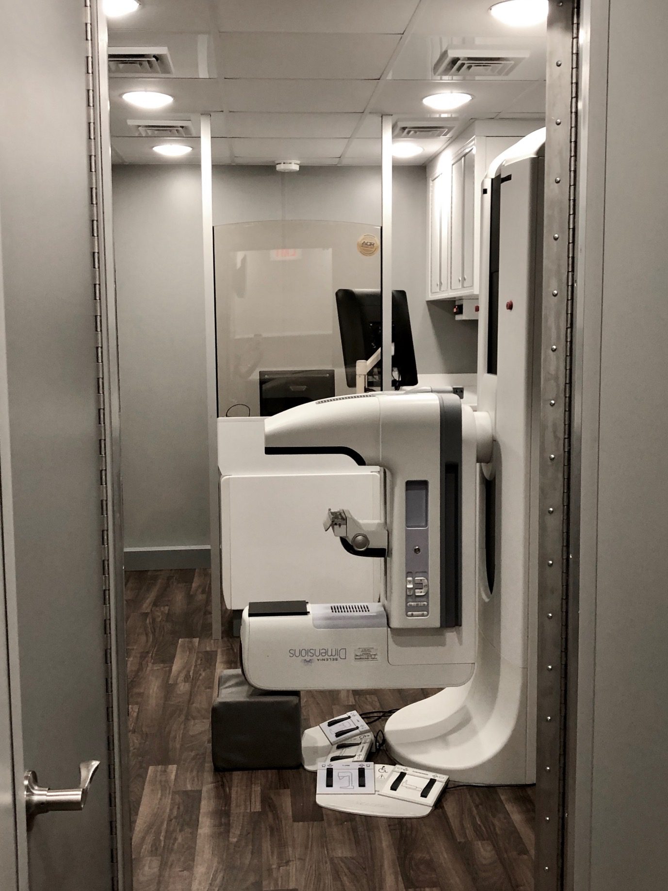
With growing demand for convenient, high-quality care, Shared Imaging delivers long-term mobile mammography solutions that meet patients where they are. Our coaches are equipped with industry-leading Hologic and GE technology, customizable for 2D or 3D screening based on your clinical needs.
THAT’S WHERE SHARED IMAGING COMES IN
While we focus on permanent, scheduled service – not interim systems – we work closely with our customers to support community engagement and planning efforts. Our mobile units deliver the same level of accuracy, privacy, and quality care patients expect from in-house services. Plus, we handle ongoing maintenance and coach aesthetics proactively, so your mobile program always reflects the excellence of your care.
WITH SHARED IMAGING, YOUR MOBILE MAMMOGRAPHY PROGRAM IS MORE THAN SERVICE – IT’S A TRUSTED EXTENSION OF YOUR PATIENT CARE.
- Flexible 2D and 3D mammography options trusted OEMs
- Mobile coaches designed for patient comfort, privacy, and high-quality care
- Help expanding access to screening and support outreach efforts that improve early detection in underserved areas
- Sustainable mobile imaging partnerships that grow with your facility
Shared Imaging’s commitment to long-term partnerships means we work closely with each customer to create dependable, customized mobile mammography solutions that align with your clinical and operational goals.
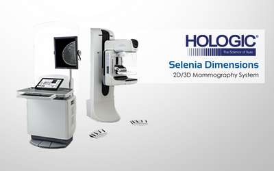
Selenia Dimensions 2D/3D Mammography System
The FIRST breast tomosynthesis technology with proven superior clinical performance to 2D mammography. Learn more about the Hologic Selenia Dimensions 2D/3D mammography system and elevate your patient care.

