Mobile PET/CT Imaging
Shared Imaging delivers advanced mobile PET/CT imaging technology solutions to healthcare facilities. These systems combine anatomical and molecular imaging for precise detection, we ensure a precise fit for your clinical needs while optimizing patient insights.

Why Bring Mobile PET/CT to Your Facility with Shared Imaging
Advanced diagnostic tools like PET/CT are essential for early detection, staging, and monitoring of serious conditions such as cancer and cardiovascular disease. However, establishing or expanding PET/CT services often involves high capital investment, staffing challenges, and logistical hurdles.
THAT’S WHERE SHARED IMAGING COMES IN
We offer custom mobile PET/CT solutions—featuring industry-leading systems from OEMs like Siemens and GE—without the need for permanent facility build-outs. Our full-service mobile imaging rental approach includes logistics, staffing, compliance, and maintenance, so your team can stay focused on patient care while we handle the rest.
With over 40 years of experience and a flexible nationwide fleet, Shared Imaging helps you:
- Launch or grow your PET/CT program without capital expense
- Serve multiple locations or rural areas on a rotating schedule
- Pilot new imaging services with minimal operational disruption
- Rely on expert support and proactive coach management
“We were in a time crunch for a mobile PET/CT and working with Shared allowed us to meet the short window to secure a unit.”
Whether you’re facing a time-sensitive need, testing market demand, or expanding access across your network, Shared Imaging is ready to help you deliver cutting-edge diagnostic imaging quickly and efficiently.
Let’s talk about how we can support your goals with the right PET/CT solution—when and where you need it.
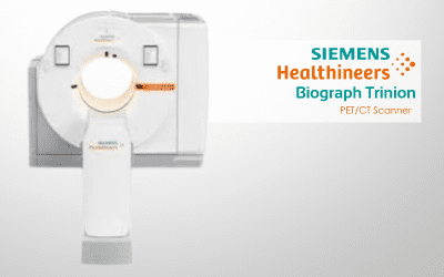
Biograph Trinion PET/CT Scanner
Integrates best-in-class hardware and software to create one high-performance platform.
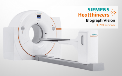
Biograph Vision PET/CT Scanner
The Biograph Vision is the next-generation of PET/CT. With its advanced capabilities and intuitive design, the Biograph Vision provides an unparalleled level of insight into patients’ conditions.
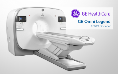
GE Omni Legend PET/CT
All-digital PET/CT system designed to deliver faster scan times, reduced dose, and advanced imaging capabilities.